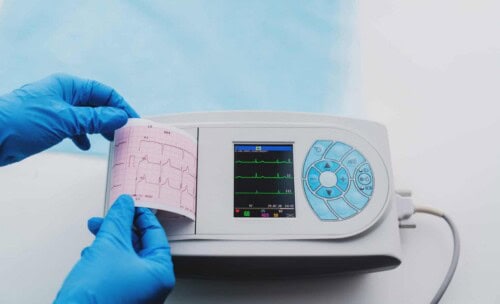
When working in a fast-paced environment, it is helpful to be able to interpret a 12-lead ECG result quickly. If you’re a clinician in urgent care or an emergency department, it’s essential that you know how to identify critical findings from an ECG. Here are some practical tips for evaluating an ECG result in a streamlined process.
Related: 12-Lead ECG Interpretation: A Primary Care Perspective
What is involved in interpreting an ECG?
Interpreting a 12-lead ECG requires a systematic approach, which includes looking at the following features:
- Rate
- Rhythm
- Intervals
- Axis
- Morphology
What is the most important thing to keep in mind when reading a 12-lead ECG?
Remember that electrical events occur in three dimensions; the length, width, and depth of the heart. To interpret electrical events using a two-dimensional ECG readout, you must use twelve different leads to visualize events in all three dimensions. A lead is the distance between two points, and each lead has both a positive and negative pole, allowing the clinician to determine in which direction the electricity is moving. Each lead provides a different point of view; you are looking at the same thing from 12 different points of view. You need to conceptualize that the readout shows you the same event from different perspectives to properly interpret the results of an ECG.
What is the first thing you look at on a 12-lead ECG?
The most important thing to review first is the rate. Abnormalities of rate can show a good amount of information, so it is a great place to start. Sinus bradycardia, or a heart rate below 60 bpm, is a common rate abnormality. It can be an indicator of digitalis, hypoxemia, hypothyroidism, hyperkalemia, and more.
Sinus tachycardia, or a resting heart rate of greater than 100 bpm, on the other hand, could be the result of caffeine, hyperthyroidism, anemia, dehydration, fever, or amphetamine use, just to name a few.
What causes rhythm abnormalities?
The next thing to review for is rhythm abnormalities, and there are many that may appear on an ECG. The most common rhythm abnormalities to look out for include heart block, atrial flutter/fibrillation, ventricular fibrillation, and monomorphic or polymorphic ventricular tachycardia (i.e. Torsade de Pointe).
What indicates heart block on a 12-lead ECG?
There are three degrees of heart block, which can all be the result of drug toxicity, electrolyte imbalances, or electrophysiological or mechanical problems. Heart block is visible on a 12-lead ECG by looking at the PR interval.
First-Degree Heart Block
First-degree heart block is characterized as a PR interval greater than 0.24 seconds, or greater than 6 mm from left to right on the ECG readout. This is typically the result of medication or electrolyte imbalances.
Second-Degree Heart Block
Second-degree heart block shows up in two ways, either Mobitz I or Mobitz II. In the case of Mobitz I, you will see a progressively longer PR interval and then a dropped beat. In the case of Mobitz II, the PR interval may be fixed with a dropped beat.
Third-Degree Heart Block
Third-degree heart block, or complete heart block, on a 12-lead ECG will show no relation between P waves and QRS. Second- and third-degree heart blocks are much more likely the result of a structural abnormality in the heart.
What causes atrial flutter or fibrillation?
Both are rhythm abnormalities that may appear on an ECG. Atrial flutter is typically a sign of hypoxemia. Atrial fibrillation may be caused by many problems, one common cause being hyperthyroidism in younger populations.
What are some common causes of ventricular fibrillation?
Many things may cause ventricular fibrillation. These include:
- Electrolyte abnormalities
- An overdose of tricyclic antidepressants
- Hypokalemia
- Hypomagnesemia
- Hypocalcemia
How can you identify Torsade de Pointe on a 12-lead ECG?
Torsade de Pointe is a form of polymorphic ventricular tachycardia visible on an ECG. It will appear as the shape of the ventricular tachycardia changes, and it will get bigger and smaller. This contrasts with monomorphic ventricular tachycardia, which always looks the same. Torsade de Pointe is commonly the result of idiopathic or drug-induced Long QT syndrome.
What is electrical axis deviation?
The normal wave of electrical activity in the heart begins in the right atria then travels down and to the left. It is important to understand the wave of depolarization, as healthy myocardial tissue will always depolarize in a predictable pattern. This will show up on an ECG as a predictable electrical axis.
What are some causes of electrical axis deviation?
Deviation can reveal one of many things that could be going wrong. With cardiac disease, you may see the wave of depolarization swing away from the area of damage or necrosis. With left ventricular hypertrophy, you will commonly see left axis deviation. This makes sense because there is more muscle mass, so it pulls the electrical activity toward that area. Similarly, with right ventricular hypertrophy, it will show up on an ECG as right axis deviation.
Other causes of axis deviation without cardiac disease are pregnancy or abdominal obesity. In both cases, the heart may have shifted in the body cavity due to the height of the diaphragm. Additionally, right axis deviation is often seen in infants, children, and adults who are tall and thin. This is because the heart is angled more up and down than normal.
What is the Quadrant Method?
For a basic ECG assessment, the quadrant method is used to quickly determine a healthy electrical axis. Using leads I and aVF (or approximated with leads I and III), you will check to see if both leads have positive QRS deflections.
On the other hand, if there is a left axis deviation, QRS in lead I is primarily positive, but in lead aVF it is primarily negative. In right axis deviation, QRS in lead I is primarily negative but QRS in aVF is primarily positive. This Quadrant Method is a visual tool for examining a 12-lead ECG and quickly identifying an assessment finding of electrical axis deviation.
What does ECG assessment of morphology include?
The final step of a 12-lead ECG interpretation is assessing the morphology. To properly assess morphology on an ECG, you need to look at each piece of the complex, which if abnormal, will tell you something different. Here is an overview of what each piece of the complex may indicate:
- P wave → atrial abnormalities
- On the right side, this would appear on an ECG as a peaked P wave of greater than 2.5 mm most commonly in leads II, III, or aVF.
- An abnormality of the left atrium would appear as P Mitraele, which shows on an ECG as a biphasic P wave in lead v1 or a notched P wave in lead II.
- QRS complex → bundle branch abnormalities
- ST segment → myocardial insult, for example, ischemia or infarction
- T wave → repolarization abnormalities
- Q waves, if present → transmural damage (shown by no depolarization in the affected tissue)
Earn CE hours with our online course on ECG Rhythm & 12-Lead ECG Interpretation Package (free with Passport Membership)!


